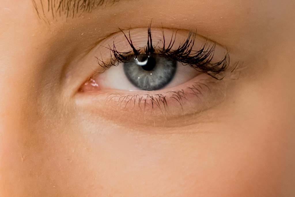
The under‑eye region is a complex landscape of delicate skin, supportive ligaments, fat pads and bone. Because of its unique anatomical features, it tends to age earlier and more dramatically than other parts of the face. For anyone considering under‑eye rejuvenation, understanding this anatomy is essential. This article explains the structures that make the area so vulnerable and explores how they change over time.
The skin: thin and poorly supported
The periorbital skin is among the thinnest on the body — up to five times thinner than facial skin¹. It contains fewer elastin and collagen fibres and has a horny layer only three cells thick compared with 15 or more elsewhere¹. There is also a scarcity of sebaceous glands in the lower eyelid². Sebum forms a protective film that reduces water loss and shields skin from environmental damage; without it, the under‑eye area is prone to dehydration and irritation. The thin dermis and lack of adipose support mean that underlying blood vessels and muscles are visible, which is why dark circles can appear blue or purple.
Constant motion and mechanical stress
The eye area is extraordinarily active. Twenty‑two muscles control blinking, smiling and squinting, leading to around 10,000 blinks per day³. These repeated micro‑contractions apply mechanical stress to thin skin, making it harder for the tissue to return to its original position. Over time, fine lines and dynamic wrinkles form. Because the skin’s ability to repair itself declines with age, repeated movements eventually create permanent creases.
Higher cellular senescence
Researchers have discovered that periorbital skin expresses higher levels of cellular senescence than other areas of the face⁴. Senescent cells no longer divide and secrete inflammatory mediators, contributing to tissue breakdown. DNA repair pathways are weaker in this region, so cumulative sun damage and oxidative stress more readily cause thinning, laxity and hyperpigmentation.
The supporting structures: ligaments, fat pads and bone
Tear trough and lid‑cheek junction: The tear trough is a natural concavity extending from the inner corner of the eye along the rim of the orbit. It lies just lateral to the anterior lacrimal crest, bounded above by orbital fat and below by the sub‑orbicularis oculi fat and cheek fat⁵. With age, bony resorption of the maxilla and orbital rim increases the hollowness of this groove⁴, while descent of the mid‑cheek accentuates the lid–cheek junction. Orbital retaining ligaments tether the skin and muscle to bone; when the soft tissue above the ligament shrinks and the bone recedes, the ligament pulls the skin inwards, creating a shadow.
Orbicularis oculi muscle and orbital septum: The orbicularis oculi is a circular muscle that closes the eyelids. Its fibres run superficially beneath the skin and deeper around the orbit. Beneath this muscle lies the orbital septum — a thin membrane separating the orbital fat from the eyelid. With age, the septum weakens and orbital fat can herniate forward, causing under‑eye “bags”⁸. In some individuals, the protruding fat sits next to the tear‑trough hollow, exacerbating the contrast between fullness and hollowness.
Sub‑orbicularis oculi fat (SOOF) and deep medial fat pad: There are two important fat compartments beneath the orbicularis muscle. The sub‑orbicularis oculi fat (SOOF) lies just below the muscle and provides mid‑facial support. Inferiorly, the deep medial fat pad fills the cheek. As we age, both compartments descend and deflate, contributing to a deepening of the tear trough and a flattening of the mid‑face. Restoring volume in these areas is therefore vital when treating under‑eye hollows; clinicians often address the cheeks before directly filling the tear trough¹⁷.
Bone resorption and facial skeleton changes: The bony orbit and maxilla undergo remodelling with age. Studies have shown that the infraorbital rim recesses, while the maxilla rotates downward and backward. These changes enlarge the orbital aperture and reduce support for the overlying soft tissues. The resulting structural hollow accentuates shadows and leads to the characteristic “skeletal eye” appearance described by oculoplastic surgeons⁷. Bone loss cannot be reversed by topical treatments; therefore volumising fillers or fat grafting often form part of rejuvenation strategies.
Why does this area age differently?
The unique combination of thin, poorly lubricated skin, continuous movement and fragile structural support means the periorbital region shows age earlier than the rest of the face. Environmental insults such as UV radiation, smoking and pollution accelerate collagen breakdown and pigmentation. The accumulation of senescent cells, higher oxidative stress and slower DNA repair in this region further exacerbate deterioration⁴. Meanwhile, bony changes and fat descent create hollows and deepen shadows, making the under‑eye area appear tired even when the rest of the face does not.
Implications for treatment
Understanding under‑eye anatomy helps explain why treatments must be tailored to individual structures:
- Skin quality – Thin skin benefits from hydration, antioxidant serums and gentle resurfacing treatments. Because there are few sebaceous glands², heavy creams may be needed to improve the barrier.
- Volume loss – Restoring cheek volume can support the lower lid and soften the tear trough¹⁷. When the hollow is very deep, carefully placed hyaluronic‑acid fillers or fat grafts can lift the depression⁴.
- Fat protrusion – Herniated orbital fat may require surgery (lower blepharoplasty) to reposition or remove the fat and tighten the septum⁸.
- Pigmentation and vascular issues – Laser treatments, chemical peels or polynucleotide mesotherapy (Lumi Eyes) may be used to improve pigmentation, microcirculation and collagen production⁴.
At Mesglo Aesthetic Clinic, practitioners assess skin thickness, ligamentous support and bone structure before recommending any intervention. A thorough understanding of anatomy ensures treatments are safe and effective, avoiding complications like overfilling or the Tyndall effect.
Conclusion
The under‑eye region is a delicate interplay of skin, muscle, fat, ligaments and bone. Its unique structural vulnerabilities explain why dark circles, fine lines and hollows appear so readily. Age‑related changes — including bone resorption, fat descent, thin dehydrated skin and higher cellular senescence — all contribute to the tired appearance many clients wish to improve. By appreciating the intricacies of under‑eye anatomy, clinicians and patients can make informed decisions about skincare, injectables, laser therapies or surgery. With a bespoke approach grounded in anatomy, it is possible to rejuvenate this challenging area while preserving natural expression and safety.

